

What Are The Different Types Of X-Rays And Their Uses?
 wholesaleXrayFilm
- 16 May, 2020
- 0 Comment
There are a huge number of conditions that would be much more difficult, if not impossible to diagnose without the use of X-rays and other imaging technologies.
From a simple toothache to a condition as complex as breast cancer, physicians and patients both would be playing a guessing game without the invention of X-rays.
Facts About X-rays
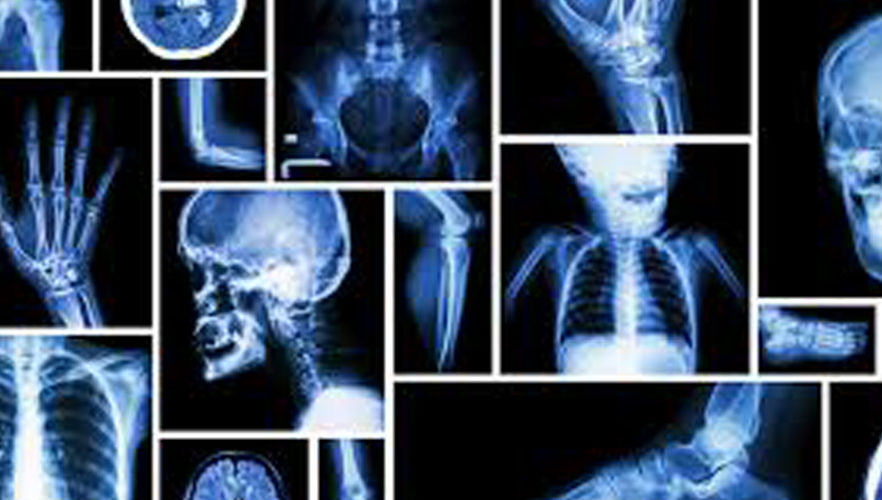
radiologist viewing xrayPhysicians are able to utilize various types of X-rays to see inside your body without resorting to an incision, and the benefits of properly administered diagnostic images far outweigh any risks. They normally are performed in a hospital radiology department, a dentist’s office, or a clinic that specializes in diagnostic procedures.
Depending on the type of X-ray being performed, you may be asked to drink a liquid or have a contrast dye injected. You may also need to fast and limit liquids, or take medications to clear out the bowels if that is the area being examined.
The 5 different types of X-rays exist because each is used for a particular reason.
Conventional Radiology
This type of traditional X-ray produces a single 2-dimensional image, which you probably have seen in an actual doctor’s office if not on a medical TV show or movie. Conventional radiology is primarily used for viewing bones, bone fractures, tissues dense in calcium, dental X-rays, and the chest.
Because they are on conventional film, the doctor and patient must wait for the results. Today’s digital images (like those produced by our digital cameras) are processed by a computer, so they can be viewed immediately. As a result, they are slowing replacing film.
CT Computerized Tomography
Computerized tomography combines a traditional X-ray with computer processing to create a better resolution. A CT scan creates a series of cross-sectional images or slices to form a 3D image. This allows Southwest Diagnostic Imaging to view different parts of the body from different angles.
A CT scan can show organs, the skeleton, tissues, and any abnormalities within these systems. This type of image can show tumors and lesions in the abdomen. A CT scan may also be used to look at the following:
• The heart to discover heart disease
• The head to find an injury, blood clots leading to a stroke, tumors, or a hemorrhage
• The lungs to discover a PE or pulmonary embolism (clots), fluid, tumors, pneumonia, or emphysema
• Bones to find fractures, bone tumors, or eroded joints
Angiography
This technique is used to examine arteries, veins, and organs in order to diagnose and treat blockages or other problems within the blood vessels. A thin tube known as a catheter is inserted into an artery or vein from the groin or arm. A contrast agent is injected into the bloodstream to make the artery or vein visible.
The goal of this test is to find blockages or narrowing of blood vessels near the heart, brain, abdomen, or legs. If a blockage is discovered, SWDIC can treat it while the angiogram is being performed.
Mammography
Most women are familiar with this special X-ray known as a mammogram. It creates detailed images of the breast to be used both as a screening tool to detect cancer at an early stage, or diagnose breast disease from symptoms like pain, a lump, or discharge from the nipple.
Tumors will show up as irregular shaped white masses. Today’s mammography has advanced to include 3D images that show the entire breast. This new technology helps to increase the likelihood of early detection of breast cancers.
Fluoroscopy
Fluoroscopy produces real time images of continuous movement within the body shown on a fluorescent screen and recorded for analysis at a later time. It shows a live image of a patient’s internal structures, and can follow the path of an injected contrast substance. A fluoroscopy test depicts real time images of a beating heart or the blood flow to the muscles of the heart.
It is also used to position a pacemaker or catheter, orthopedic implants during a surgical procedure, or to view contrast agents. This is also the same technology used during a barium enema to view movement through the gastrointestinal tract.
These 5 types of X-rays are a valuable diagnostic tool for physicians that help catch diseases faster and help patients live longer! Consult with Southwest Diagnostic Imaging if you have questions about an upcoming X-ray.